Anatomy Of Musckes Sndctendons : Two-Jointed Muscles of the Lower Body: What They Are and ... / There are four muscles that comprise the muscles of mastication.
Anatomy Of Musckes Sndctendons : Two-Jointed Muscles of the Lower Body: What They Are and ... / There are four muscles that comprise the muscles of mastication.. The primary function of the knee is to hinge at the lower extremity. There's no strict demarcation or dividing line between the tendon and the covering around this muscle but that covering is called is called the epimysium fp my cm and it's really just connective tissue that covers the muscle kind of protects it reduces friction. Muscular contraction is necessary for voluntary and involuntary movement of limbs, stabilization of joints, maintaining luminal diameter (in the case of arteries, bowel, etc), and to produce heat. Learn about human anatomy muscles with free interactive flashcards. Smooth muscles are found in the walls of many organs, such as the stomach and in blood vessels.
The muscles of the torso, examined in the previous chapter, include a few that attach directly into the upper arm and help move the humerus at the shoulder joint. Muscles of mastication are classified as main and accessory muscles. It elevates and protrudes the mandible. Muscular contraction is necessary for voluntary and involuntary movement of limbs, stabilization of joints, maintaining luminal diameter (in the case of arteries, bowel, etc), and to produce heat. Inflammation of this region caused by repetitive stress or trauma may lead to pain and numbness known as carpal tunnel syndrome.
Pin by ashlee brown on Anatomy | Leg muscles anatomy ... from i.pinimg.com Anatomy of a muscle cell. In the diagrams below, i'll be showing muscle groups in color, with a black line to show the forms that would show through the skin (i also show protruding bones that would do the same). Movement of the mandible at the temporomandibular joint). These muscles originate from the surface of the skull and insert onto the mandible.¹. Smooth muscle contractions are involuntary movements triggered by. This handbook of general anatomy has been written to meet the requirements of students who are newly admitted to medica. Learn about human anatomy muscles with free interactive flashcards. The muscular system is responsible for the movement of the human body.
There's no strict demarcation or dividing line between the tendon and the covering around this muscle but that covering is called is called the epimysium fp my cm and it's really just connective tissue that covers the muscle kind of protects it reduces friction.
In this section, learn more about the anatomy of the muscles of the neck. They are associated with muscles discussed in the section above (see. This handbook of general anatomy has been written to meet the requirements of students who are newly admitted to medica. Through a simple and intuitive interface it is possible to observe systems: Learn about the muscles, tendons, bones, and ligaments that comprise the knee joint anatomy. These muscles originate from the surface of the skull and insert onto the mandible.¹. Topographically, the muscles in this group are classed along with the lateral torso wall and upper extremity, which is due to their location as well as their genetic development based on their embryological origin. By contracting, they also aid the venous return of blood to the heart and with age, these components of the musculoskeletal system progressively degenerate, which contributes to frailty and increases the risk of falls and fractures. Circular skeletal muscles are made up of fibers that are arranged in a circular manner. Convergent muscles contain fibers that have a wide origin, but converge in order to attach to a narrow tendon. Muscles of mastication are classified as main and accessory muscles. Want to learn more about it? The tendons of these muscles pass through a small corridor in the wrist known as the carpal tunnel.
The muscles of the torso, examined in the previous chapter, include a few that attach directly into the upper arm and help move the humerus at the shoulder joint. Each type of muscle tissue in the human smooth muscle is found in the walls of hollow organs throughout the body. This is a table of skeletal muscles of the human anatomy. Inflammation of this region caused by repetitive stress or trauma may lead to pain and numbness known as carpal tunnel syndrome. Convergent muscles contain fibers that have a wide origin, but converge in order to attach to a narrow tendon.
Neck Muscles Anatomy - Posterior Triangle, Prevertebral ... from i1.ytimg.com Circular skeletal muscles are made up of fibers that are arranged in a circular manner. You can click the links in the image, or the links below the image to find out more information on any muscle group. Anatomical terms structures of the knee bones of the knee ligaments in the knee cartilage of the knee muscles around the knee tendons in the there are numerous tendons around the knee that also help to stabilize the knee. Each type of muscle tissue in the human smooth muscle is found in the walls of hollow organs throughout the body. Smooth muscle contractions are involuntary movements triggered by. Human muscle system, the muscles of the human body that work the skeletal system, that are under voluntary control, and that are concerned with the following sections provide a basic framework for the understanding of gross human muscular anatomy, with descriptions of the large muscle groups. Musculoskeletal, cardiovascular, nervous, respiratory, digestive, urogenital (male and female), endocrine, lymphatic, eye and ear. Anatomy of the short head of the biceps brachii muscle.
Learn about human anatomy muscles with free interactive flashcards.
• the muscular system develops from intra embryonic mesoderm. This is a table of skeletal muscles of the human anatomy. There's no strict demarcation or dividing line between the tendon and the covering around this muscle but that covering is called is called the epimysium fp my cm and it's really just connective tissue that covers the muscle kind of protects it reduces friction. In this section, learn more about the anatomy of the muscles of the neck. Knee function is determined in large part by the anatomy of the joint. The primary function of the knee is to hinge at the lower extremity. Topographically, the muscles in this group are classed along with the lateral torso wall and upper extremity, which is due to their location as well as their genetic development based on their embryological origin. Want to learn more about it? The anterior and middle scalenes originate from the transverse processes of certain cervical vertebrae and attach to the first rib. Anatomy 3d atlas allows you to study human anatomy in an easy and interactive way. Smooth muscle contractions are involuntary movements triggered by. In the muscular system, muscle tissue is categorized into three distinct types: Muscles of mastication are classified as main and accessory muscles.
Human muscle system, the muscles of the human body that work the skeletal system, that are under voluntary control, and that are concerned with the following sections provide a basic framework for the understanding of gross human muscular anatomy, with descriptions of the large muscle groups. Smooth muscles are found in the walls of many organs, such as the stomach and in blood vessels. Learn about the muscles, tendons, bones, and ligaments that comprise the knee joint anatomy. Inflammation of this region caused by repetitive stress or trauma may lead to pain and numbness known as carpal tunnel syndrome. In this section, learn more about the anatomy of the muscles of the neck.
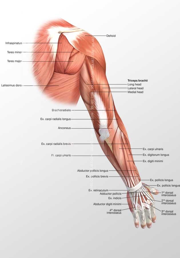
Muscle 3D Illustrations|human body illustrations from www.3dlabz.com There are around 650 skeletal muscles within the typical human body. Anatomical terms structures of the knee bones of the knee ligaments in the knee cartilage of the knee muscles around the knee tendons in the there are numerous tendons around the knee that also help to stabilize the knee. The muscular system is responsible for the movement of the human body. Muscles of mastication are classified as main and accessory muscles. Topographically, the muscles in this group are classed along with the lateral torso wall and upper extremity, which is due to their location as well as their genetic development based on their embryological origin. This handbook of general anatomy has been written to meet the requirements of students who are newly admitted to medica. Each of these muscles is a discrete organ constructed of skeletal muscle tissue, blood vessels, tendons, and nerves. Learning to draw muscles may conjure medical charts in daunting details, but such complexity is unnecessary.
Anatomy 3d atlas allows you to study human anatomy in an easy and interactive way.
By contracting, they also aid the venous return of blood to the heart and with age, these components of the musculoskeletal system progressively degenerate, which contributes to frailty and increases the risk of falls and fractures. Inflammation of this region caused by repetitive stress or trauma may lead to pain and numbness known as carpal tunnel syndrome. You can click the links in the image, or the links below the image to find out more information on any muscle group. Each of these muscles is a discrete organ constructed of skeletal muscle tissue, blood vessels, tendons, and nerves. • definitions • introduction • development of muscles • classification • anatomy of skeletal muscle • muscle physiology • properties • muscles of development of muscles. It elevates and protrudes the mandible. Smooth muscles are found in the walls of many organs, such as the stomach and in blood vessels. This handbook of general anatomy has been written to meet the requirements of students who are newly admitted to medica. The muscles of mastication are a group of muscles responsible for chewing (i.e. Learn about the muscles, tendons, bones, and ligaments that comprise the knee joint anatomy. The muscles of the torso, examined in the previous chapter, include a few that attach directly into the upper arm and help move the humerus at the shoulder joint. The tendons of these muscles pass through a small corridor in the wrist known as the carpal tunnel. Learning to draw muscles may conjure medical charts in daunting details, but such complexity is unnecessary.


0 Komentar